
 By
By Scientists have made a remarkable discovery about one of the most complex organs in the human body: The brain appears to begin life as two separate nervous systems that are ultimately brought together inside the skull.
The Stanford Medicine-led study, published Friday in Nature Neuroscience, found that the front and back of the brain develop from entirely different populations of early cells. The discovery overturns a longstanding assumption that the entire brain traces back to a single developmental starting point and could give researchers new tools for studying devastating neurological diseases.
“We’ve shown for the first time that the front of the brain arises from a totally different progenitor cell than the back of the brain,” said Kyle Loh, PhD, associate professor of developmental biology and senior author of the study. “Our discovery means that we can now grow neurons from the back of the brain, the hindbrain, in a petri dish and study their functions.”
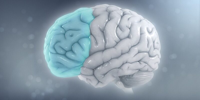
The adult brain is generally divided into the forebrain, midbrain and hindbrain. The forebrain handles many of the abilities most associated with human thought, including language, consciousness and abstract reasoning. The hindbrain, or brainstem, performs some of the body’s most essential jobs, regulating breathing, heartbeat, sleep and hunger while also controlling muscles used for swallowing, facial movement and speech.
Until now, scientists largely believed those regions developed from the same early progenitor cells. The Stanford researchers instead traced the brain’s origins to gastrulation, an early stage of embryonic development when the basic structure of the body begins to form.
Graduate students Carolyn Dundes and Rayyan Jokhai, co-first authors of the study, found two distinct populations of progenitor cells in mouse embryos. Cells expressing the gene Otx2 became the forebrain and midbrain, while cells expressing Gbx2 developed into the hindbrain. The two populations did not overlap.
Researchers also found differences in the cells’ chromatin, the material that helps determine which genes can be activated. From the earliest stages of development, the front and back portions of the nervous system appeared to be following separate biological paths.
“Previous attempts to make hindbrain neurons likely tried to coax forebrain and midbrain progenitors into hindbrain cells, which our study shows is not possible,” Jokhai said.
“In stem cell biology, people are always fixated with creating the end cell type, like the neuron,” Jokhai said. “But it’s important to begin at the earliest stages of embryonic development. Our careful attention to that early time point allowed us to find this fundamental split in brain development.”
That discovery helped the scientists accomplish something with major potential for medical research. By following the correct developmental pathway, the team successfully turned human pluripotent stem cells into functional hindbrain motor neurons in the laboratory. The cells fired electrical signals and produced proteins associated with parts of the brainstem that control facial and swallowing muscles.
The advance could open new possibilities for studying spinal muscular atrophy, or SMA, and amyotrophic lateral sclerosis, commonly known as ALS. Both diseases can damage hindbrain neurons involved in swallowing and breathing, but researchers cannot simply take brainstem tissue from living patients to study what is going wrong.
Growing those neurons in a laboratory could allow scientists to watch the disease process unfold at the cellular level and potentially test new treatments.
The discovery also revealed something extraordinary about the deep history of the human brain. Researchers found the same two-origin pattern in chickens, zebrafish and acorn worms, animals whose evolutionary paths separated from humans more than 550 million years ago. Jellyfish, which diverged even earlier, have two nervous systems located at opposite ends of their bodies.
“Our research suggests that evolution took two existing neural systems and pushed them together spatially,” Loh said. “Having the brain as one organ would probably be more efficient, but we rely on this primordial way to make the brain as two separate pieces.”
“I was surprised at our findings because the word ‘brain’ implies a contiguous organ that likely has a singular origin,” Jokhai said. “But even 500 million years ago, there were these separate neural systems, which now almost operate as one, which is very cool.”
The implications could extend beyond diseases such as SMA and ALS. The hindbrain also contains circuits involved in hunger that are targeted by drugs including semaglutide, raising the possibility that the new laboratory models could help researchers better understand other areas of human health.
The Stanford team now plans to investigate the developmental origins of the spinal cord and study more closely how SMA and ALS damage hindbrain neurons.
“Now we have a model to better understand these devastating diseases, and work toward regenerative therapies for them,” Jokhai said. “This is a very exciting new frontier in brain research.”
It looks like the textbooks will have to be rewritten.